Logo Eara

Brain

AuthorEARA team
Brain research is one of the areas of biomedical science where animal studies are most commonly used, reflecting the brain’s complexity and its close integration with the rest of the body. While non-animal approaches are advancing, many aspects of brain function, disease progression and behaviour can currently only be studied in living organisms. 
Brain research, the field of research where most animals are used, stands at a critical juncture where animal studies remain essential for understanding the functions of the brain, both in basic research and drug testing. The brain's vast complexity — connecting all organs and systems while remaining our least understood organ — requires integrated biological systems that only living organisms can provide. Current treatments for brain disorders often target non-specific mechanisms with significant side effects, highlighting major knowledge gaps in our understanding of neural circuits, disease progression and therapeutic interventions.
One of the greatest challenges in neuroscience research is tackling neurodegenerative diseases such as dementia and Parkinson's disease, which currently affect tens of millions of people across Europe. As the proportion of the elderly in Europe increases, it is vital that the most effective methods of research are used to combat this challenge. It is estimated that brain disorders may cost as much as 45% of Europe's annual health budget (800bn euros and large-scale brain research is now underway, investing billions of dollars and euros, to fund the Human Brain Project in Europe and the Brain Activity Map Project (BRAIN Initiative) in the USA.  
At present, society’s best hope of finding drugs and other treatments for diseases of the brain relies on research using animals. While non-animal methods of study have made progress in some fields of biomedical research, their use in neuroscience remains extremely limited due to the complex and interconnected structure of the brain. In most cases, a living and behaving organism remains the only viablemodel to study the brain in action.
The European Brain Council, which represents scientific societies, patient organisations, professional societies and industry partners, recently expressed its support for the continued use of animals in neuroscience research.  

Why are animals used in brain research? 

Media Bank Image
Understanding the brain requires studying both healthy function and disease states in living, behaving organisms.  
Basic research
Over the past 50 years, basic research using animals has dramatically improved our understanding of the brain and the nervous system, winning several Nobel Prizes for Physiology and Medicine, including: 
  • In 2000, for the discovery of dopamine that transmits signals in the brain and is involved in diseases such as Parkinson's, using animals including mice and rats. 
  • In 2013, for understanding how proteins are transported in cells, using hamsters, mice and rats. 
  • In 2014, for the discovery of cells involved in a 'positioning' system in the brain, using rats. 
Several mechanisms and brain functions are still unknown, including those that govern human and animal behaviour, reflexes and cognition. Scientists at Massachusetts Institute of Technology (MIT) have discovered how the brain can recover from a distraction to return to the task at hand, in a study using monkeys, which may help explain how the brain responds to memory challenges, findings which could also apply to humans.   
Modelling disease mechanisms 
Animals allow researchers to observe how disease develops over time, from initial molecular changes through symptom emergence. Genetically modified mice can carry mutations found in human patients, enabling study of specific disease pathways. Larger animals like monkeys provide models where brain structure and organisation closely parallel humans. 
Testing interventions 
Before any treatment reaches human trials, it must demonstrate safety and efficacy in living systems. Animals enable testing of drugs, gene therapies and devices like brain implants in complete organisms where interactions between the nervous system, immune system and other organs can be evaluated. 
Investigating behaviour and cognition 
Many brain conditions affect memory, emotion, movement or sensation. Animals can perform behavioural tasks that reveal how specific brain regions or circuits contribute to these functions and what changes when disease occurs. 
Understanding brain development 
Studying how brains forms and changes from the embryo through ageing requires animals with observable development and shorter lifespans than humans, allowing researchers to examine critical periods and long-term effects. 

Which brain diseases benefit from animal studies?

Brain tumours, epilepsy, sleep disorders and other neurological conditions represent diverse challenges where animal research continues providing crucial insights. 
For instance, animal research can provide insights into both how brain damage can occur and how to stop it. A group at the Institute of Science and Technology Austria found that starving the brain of certain dietary nutrients can impact brain development in mice, while in zebrafish, a team at Ludwig Maximillian University, Germany, were able to prevent the damage caused by brain scarring. Scientists activated existing immune cells in the brain called microglia, to study their effects – microglia are key to forming scar tissue. 
Epilepsy research at Stanford University in the USA achieved a significant milestone by successfully switching brain cells 'on and off' within mouse brains, preventing seizures using optogenetics. This precision approach, impossible without intact neural circuits in living animals, offers hope for more targeted treatments with fewer side effects than current medications. 
Brain tumours present particular treatment challenges due to their location and aggressive growth. A study in mice at EARA member the University of Zurich, Switzerland, has allowed for the testing and refinement of treatments for aggressive brain tumours by showing that a protein-antibody combination treatment can slow or reverse the growth of cancer cells. Another study at the Hospital for Sick Children, US, and EARA member University of Toronto, Canada, used gene editing in mice to discover why childhood brain tumours resist radiation therapy, identifying genes that could be targeted to restore treatment effectiveness. 
The link between the nervous system and digestive system has been investigated in mice, for example at EARA member the Champalimaud Foundation, Portugal, which looked at feeding behaviour in mice to provide insights into understanding and treatingobesity, demonstrating how neurological research addresses conditions not traditionally viewed as brain disorders. 
Sleep disorders often connect to other brain conditions, as shown in research at the University of Queensland, Australia, which found that a condition called sleep apnoea was associated with a higher risk of developing Alzheimer’s disease. ​ 
The brain and spinal cord also have an important role in sexual behaviour and premature ejaculation. Researchers at EARA member Champalimaud Centre for the Unknown, Portugal, and University of Bordeaux, France, discovered that specific spinal cord neurones and a type of brain cells control ejaculation and arousal in mice and rats, suggesting that rodents may be a good model to study premature ejaculation and human arousal regulation. 

In the absence of scientifically valid methods that can replace particular animal procedures, phasing out the use of animals in medical research would have major consequences and impact the quest to improve the quality of life of the many citizens affected by brain conditions, neurological and mental alike.

European Brain Council, statement on animal research, 2023

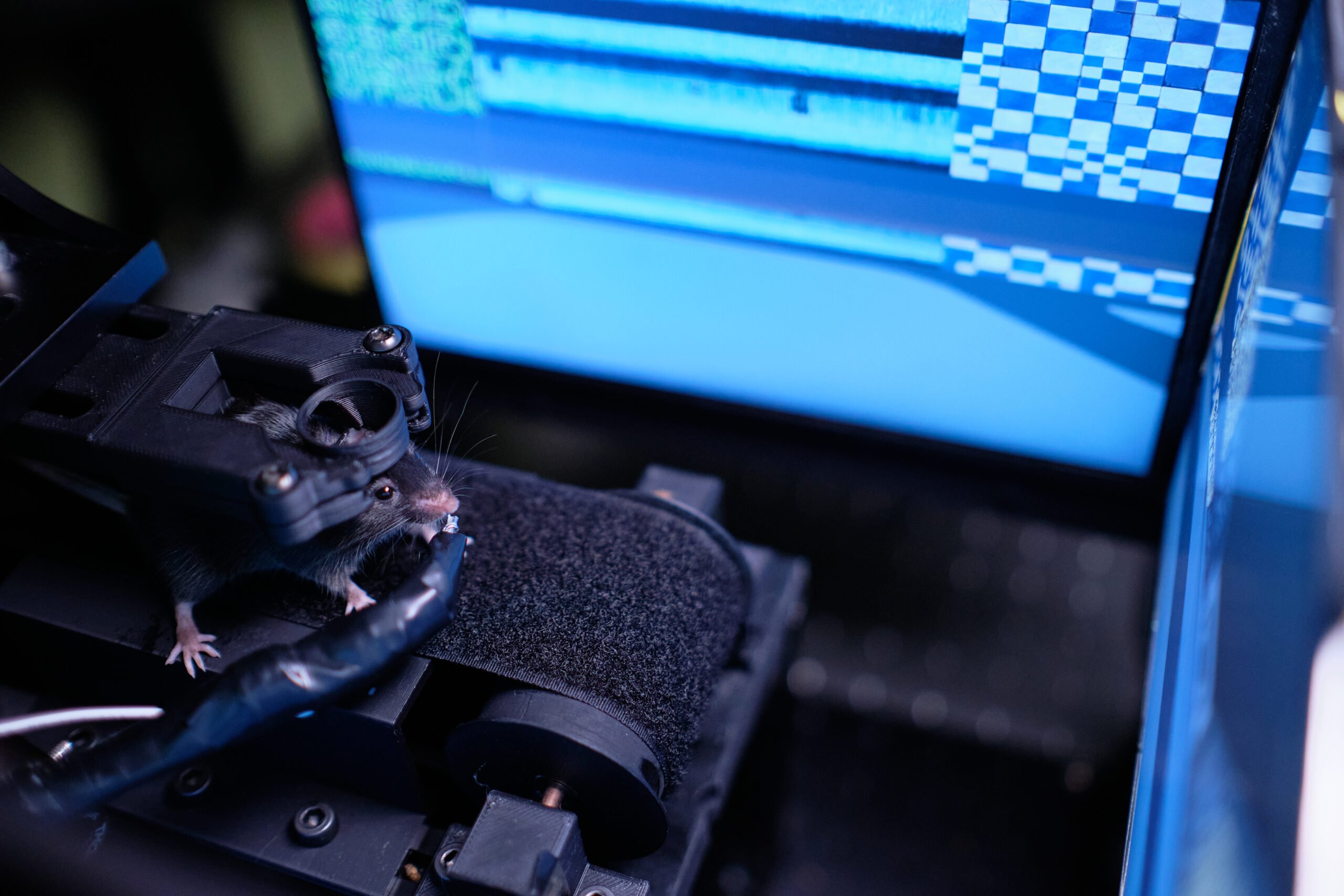
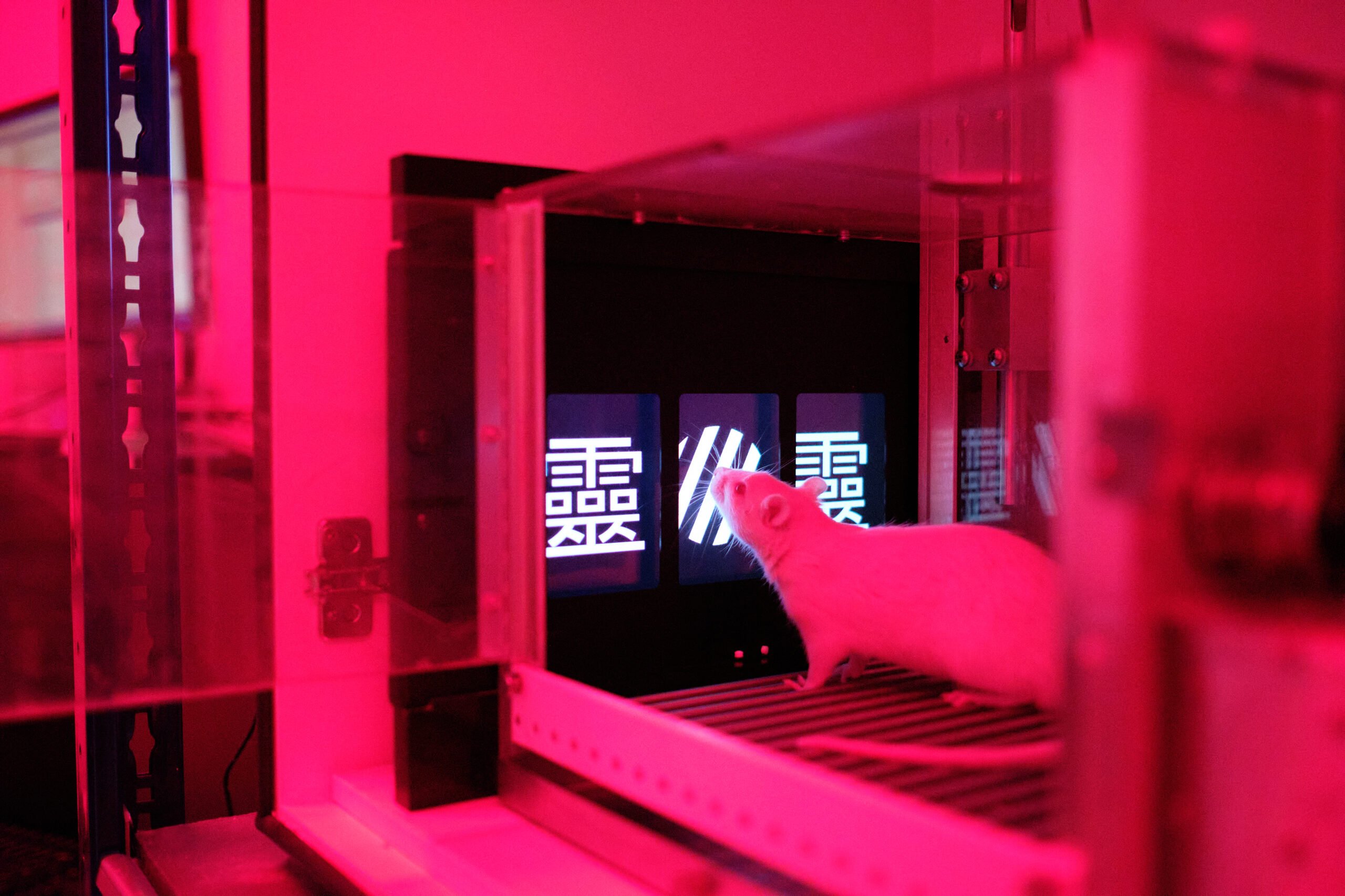
Which animals are used in brain research?

The use of animals in all areas of biomedical and scientific research is tightly regulated in the EU and many other parts of the world, to ensure that animal research only takes place when there is sound scientific justification and no other viable method is available.  
A high standard of animal welfare benefits not only the animal, so that they are kept in an environment that minimises stress and, where possible, can mimic natural conditions, but also to maximise reliable results in studies.  
All researchers who use animals must follow the principles of the 3Rs (replacement, reduction, refinement), while alternative methods to complement animal research are increasingly being developed. However, for brain and neuroscience research, such alternatives are still very limited compared to other research areas and are not yet able to mimic the complexities of living brains.  
Media Bank Image

Having such a close genetic relationship to humans, monkeys are one of the most valuable animal models used in research. Less than 0.2 per cent of the animals used in research in the EU are monkeys, however their impact in providing the most reliable information for what is happening, or what is going to happen, in humans cannot be underestimated.  
​All animal experiments are strictly regulated and reviewed by ethical committees before they are allowed to proceed and the use of monkeys in research is only permitted when there is no other animal or non-animal model that could provide the same answer. In addition, in Europe, research with great apes such as chimpanzees – the animal that is the most closely-related to humans – is prohibited. 
While there are understandable ethical worries about using monkeys, they continue to be an essential model for studying the function of the brain due to the similarity in structure and composition with humans. The prefrontal cortex structure in non-human primates more closely resembles humans than does the rodent brain, making monkeys essential for studying complex behaviour, emotion, vision and higher cognitive functions. 
Much of what we know today about complex behaviour and emotion, vision and higher cognitive function has been gained from the study of monkeys – as well as insights into how to treat these functions when they go wrong, such as in vision loss, paralysis and stroke.  
An international task force published a position paper in NPJ Parkinson's Disease highlighting how non-human primates remain critical for Parkinson's research and understanding ageing. Monkey studies have enabled the mapping of brain circuits and improved understanding of deep brain stimulation, translating directly into patient therapies. Their genetic, anatomical and behavioural similarities to humans make them irreplaceable for treatment development. 

"Lots of neurological ph lifetime, which is more difficult to achieve in larger animals and humans. For example, research at the Institute for Research in Biomedicine and SJD Paediatric Cancer Center Barcelona, both Spain, engineered fruit flies to express a variant of a cancer gene that causes a rare bone cancer called Ewing sarcoma, which has a survival rate of only just over 50% in teenagers, and less than 30% after the cancer has spread to other parts of the body. This resulted in the same tumour development as seen in humans, and was the first time the disease was modelled in an animal, after failed attempts to do so in mice. 

The causes of the rare neurodevelopmental disorders, Harel-Yoon syndrome and Yoon-Bellen syndrome, were both identified as due to specific genetic mutations thanks to studying fruit flies, by researchers at Baylor College of Medicine, USA. Although this gene mutation had been linked to neurological symptoms in humans, it was only by determining the effect in fruit flies that researchers could confirm the cause – paving the way to targeted treatments. 

Meanwhile, in a fruit fly study at the University of Sydney, Australia, researchers were able to pinpoint how a mutation underpinned a key mechanism behind episodic ataxia, a condition that severely affects balance and co-ordination. 

Fruit flies reach adulthood in just two weeks, so using them in research opens doors to exploring the long-term effects of a disease and how it progresses over a lifetime, which is more difficult to achieve in larger animals and humans. For example, research at the Institute for Research in Biomedicine and SJD Paediatric Cancer Center Barcelona, both Spain, engineered fruit flies to express a variant of a cancer gene that causes a rare bone cancer called Ewing sarcoma, which has a survival rate of only just over 50% in teenagers, and less than 30% after the cancer has spread to other parts of the body. This resulted in the same tumour development as seen in humans, and was the first time the disease was modelled in an animal, after failed attempts to do so in mice. 


Limitations of animal models 

While animal models have contributed to major progress in neuroscience, they have inherent limitations that researchers must acknowledge and address. No animal species perfectly replicates human brain structure, functionor disease. Mice and rats, despite their widespread use, have different brain organisation than primates, particularly in regions governing higher cognitive functions. Disease progression timelines differ, with conditions developing over decades in humans potentially manifesting in months in rodents. 
Behavioural assessments face challenges - animals cannot report subjective experiences like pain intensity or emotional states, requiring researchers to infer these from observable behaviours that may not directly correspond to human experiences. Genetic models carrying human disease mutations may not develop identical symptoms, as other genetic and environmental factors influence outcomes. 
These limitations don't invalidate animal research but require careful interpretation of results, use of multiple complementary models and rigorous validation before translating findings to humans. Researchers continuously work to refine animal models and develop better predictive methods while maintaining transparency about what animal studies can and cannot tell us. 

New approach methodologies in brain research 

New approach methodologies, such as advanced cell cultures, organoids and computational models, complement animal studies in neuroscience and are making remarkable progress. The EU Commission Joint Research Centre has published a collection of over 550 new approach methodologies for studying neurodegenerative diseases. These approaches offer new ways to study human brain cells directly and are contributing to earlier-stage discovery, particularly in understanding molecular mechanisms and screening potential treatments. 
Brain organoids are 3D cultures of different cell types grown from human stem cells that aim to reproduce some aspects of brain development and organisation. They allow researchers to study human brain cells in ways that are not possible in living people and can be used to investigate early developmental processes or test drug effects in a human cellular context. At Johns Hopkins University, US, researchers developed the first multi-region brain organoid by fusing nerve cells from different brain regions using proteins that act like biological "superglue." As tissues grew together, they formed connections and began responding as a network. Though much smaller than actual brains, containing six to seven million neurones versus tens of billions in adults, these organoids show promise for studying how complex brain diseases like schizophrenia, autism and Alzheimer's affect the whole brain, not isolated regions. 
At the same time, organoids remain simplified systems. They do not yet reproduce the full complexity of the human brain and lack key physiological features such as blood vessels, immune cells and sensory inputs. Researchers at EPFL and the Roche Institute of Human Biology, Switzerland, have highlighted that, without blood vessels or interactions with the immune system, organoids cannot fully mimic processes such as neuroinflammation, brain ageing or responses to injury. This limits their ability to capture diseases that unfold over long periods or involve interactions between the brain, other organs and the environment. Currently, researchers are still at the first stages of developing organoids with blood vessels. However, recent research has made progress in the development of more complex organoids. Researchers at Stanford Medicine have generated lab-grown human heart and liver organoids that form their own blood vessels. However, recent research has made progress in the development of more complex organoids. Researchers at Stanford Medicine, US, have developed lab-grown human heart and liver organoids that form their own blood vessels.

Organoids don’t let us model the biology of entire organisms. Some questions can only be answered by examining an actual organism – an animal. It will take time before more realistic models are developed that will mark a major step towards replacement.

Matthias Lütolf, professor at EPFL and co-founder of the Roche Institute of Human Biology
recent review highlighted that brain organoids used to model Alzheimer’s disease typically lack non-neuronal brain cells, such as microglia and oligodendrocytes, meaning they cannot reproduce essential features such as brain inflammation, myelin loss or blood–brain barrier dysfunction that are central to the disease. The review stresses that heterogeneity in organoid size, composition and structure can lead to inconsistent Alzheimer’s-related characteristics, making it difficult to reliably interpret results without validation in more integrated in vivo models.The authors also note that, even after lengthy culture periods, organoids remain immature and therefore struggle to model age-related neurodegeneration, which is a major limitation given that ageing is the strongest risk factor for Alzheimer’s disease.  
Nonetheless, current research is underway to make organoids more reliable for ageing studies, including the use of organoids derived from patient’ stem cells to uncover new biomarkers, predict adverse outcomes and study newtreatments of ageing-related diseases. Despite these advances, organoids still have significant limitations as a method to study ageing. The fundamental challenge lies in recapitulating the decades-long process of biological ageing in laboratory conditions. Current organoids cannot replicate the cumulative effects of chronic inflammation and accumulation of cellular damage, nor capture the systematic interactions between organ systems that characterise natural ageing. They also fail to capture how the environment changes can shape age-related pathology in living organisms. 
Advances in stem cell biology have also expanded the range of brain cell types that can be studied in the laboratory.Scientists at ETH Zurich, Switzerland, were for the first time able to grow over 400 different types of human nerve cells from human stem cells, a dramatic increase from the few dozen types previously achievable. By treating cells with specific combinations of morphogens (molecules that guide cell identity during development), researchers created nerve cells from different brain regions with distinct functions, such as sensing pain, cold or movement. Comparing lab-grown cells with human brain cell databases confirmed their similarity to actual brain cells, greatly expanding the nerve cell types available for laboratory research. the nerve cell types available for laboratory research. 
Researchers at Stanford University, USA, were able to transplant human brain organoids into rats and showed the different cell types had become integrated in the animals’ brain. This paves the way for an improved understanding of brain development and disorders, although such developments also bring about new ethical issues.  
Organs-on-a-chip are advancing rapidly as a strategy to test human cells from different physiological systems in a more integrated way. However, organ-on-a-chip platforms still lack a universal “blood mimetic” capable of sustaining multiple cell types, making it difficult to recreate truly physiologically relevant conditions in complex multi-organ chips. 
NAMs are contributing to the reduction and replacement of animal use in specific contexts, particularly for screening and mechanistic studies. At the same time, developing accurate and predictive alternatives depends onunderstanding how the brain works, knowledge that continues to come largely from animal research. Therefore,neuroscience advances, including in new approach methodologies, continue to come largely from animal research.Using each model where it is most appropriate and informative, while working toward future technologies, may gradually reduce the need for animals. 

We shouldn’t forget that organoids are possible thanks only to the massive amount of research that came before. Many of the questions and concepts we’re studying in developmental biology were first explored in flies and mice, for example. These types of models can’t be replaced so easily.

Fides Zenk, researcher at EPFL
Other useful sources
Webinar on “Brain Research Needs Animal Models: Let’s Talk About It!”, Federation of European Neuroscience Societies
The continued need for animals to advance brain research, Neuron
Understanding Brain Research Models, Dana Foundation