
Researchers in Canada have discovered that blood flows differently in the mouse brain when responding to touch and pain, challenging a premise of medical brain imaging in neurological diseases and potentially improving how brain scans are interpreted.
Functional magnetic resonance imaging (fMRI) is a non-invasive medical exam used to measure brain activity. However, it does not measure the activity of the neurones directly but changes in oxygen in the blood associated with brain activity.
Researchers from EARA member University of Montreal wanted to check if the blood flow is similar when the brain is responding to different stimuli, which is assumed when interpreting fMRI images of the brain.
Using high-resolution microscopy, they observed the blood flow in the cortex – the outer layer of the brain – while mice received touch or pain stimuli. The pain stimuli were induced by shining light on the hind paws of mice whose pain receptor neurones had been genetically modified to respond to light. The researchers found that while the average flow in the cortex was similar under both conditions, blood flow at different depths within the cortex was reduced by more than 50% when mice were feeling pain compared with touch. By analysing the images using computational models, the team discovered that these differences occur because the brain activity associated with pain and touch directs blood flow through distinct networks of blood vessels with different diameters.
“We need to start viewing blood vessels as an active architecture of neural circuits, not just a delivery system,” said Ravi Rungta, from the University of Montreal and leader of the study published in Science.
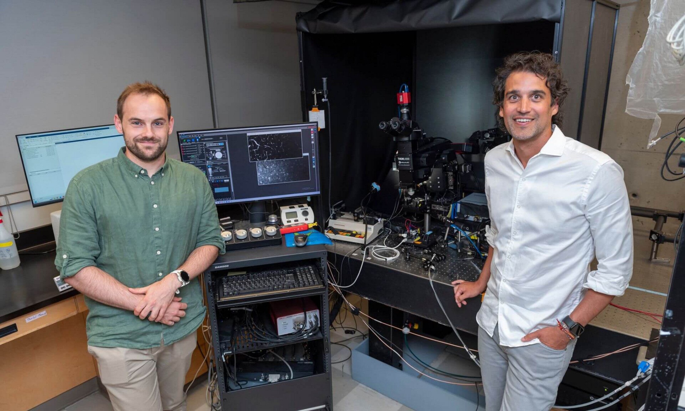
Researchers Antoine Malescot and Ravi . CREDIT: AMÉLIE PHILIBERT, UNIVERSITÉ DE MONTRÉAL