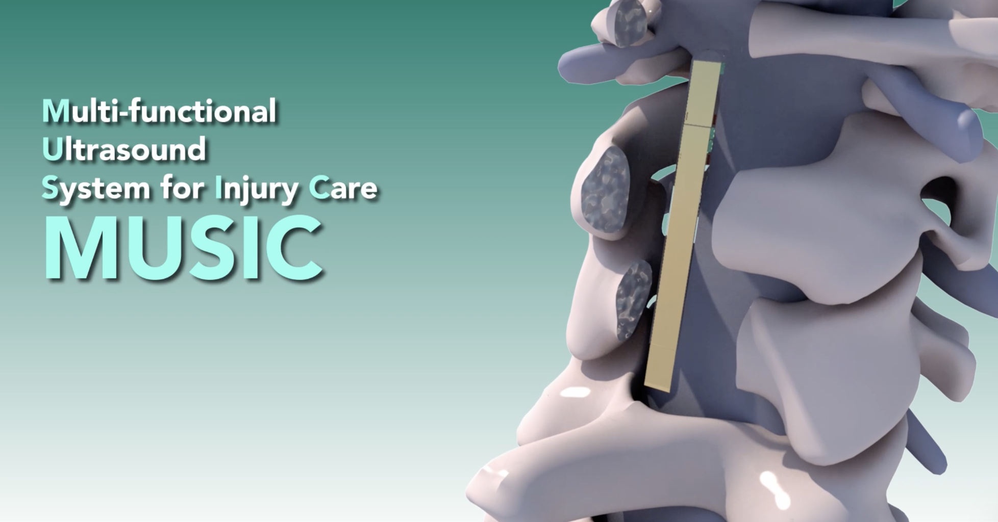
Researchers in the US have tested a miniature ultrasound device in pigs that provides real-time images and blood-flow information near spinal cord injuries, which could aid clinicians to detect complications and adapt patient treatment during recovery.
Spinal cord injuries can disrupt communication between the brain and body, causing lasting changes in movement, sensation and other bodily functions. Changes in blood circulation and fluid accumulation can also contribute to further tissue damage.
Researchers at Johns Hopkins University, Maryland, developed the Multimodal Ultrasound System for Injury Care (MUSIC), a small device designed to sit over the spinal cord. It combines diagnostic ultrasound, which uses sound waves to produce images and measure blood flow, with focused ultrasound, which could be used to regulate blood flow by concentrating sound energy on a specific area.
In anaesthetised pigs, the researchers surgically induced spinal cord injuries and temporarily placed MUSIC over the spinal cord. They demonstrated that the device provided real-time images, detected bleeding and monitored blood flow around the injured area. In non-injured pigs, using MUSIC’s focused ultrasound function, the researchers were able to change blood flow velocity.
“This research represents a fusion of multiple industries, disciplines and applications that we envision has the power to fine-tune and personalize the care that a person receives following their surgery, which could support and improve their health outcomes months and years later,” said Nicholas Theodore, professor at Johns Hopkins University and author of the study published in Science Advances.
The team hopes the device, which would be implanted in patients already requiring surgery, could allow medical staff to monitor patients in intensive care without repeatedly moving them for conventional imaging. Further research is needed to assess its safety and therapeutic capabilities before it can be considered for use in patients.