A study in Germany has created the first detailed map of the proteins that are involved in initiating pain in neurones, helping to identify potential therapeutic approaches for chronic pain.
Researchers from the Helmholtz Centre for Infection Research, in collaboration with the Max Delbrück Center (MDC), an EARA member, analysed how mice pain neurones, called nociceptor, responded to the molecule NGF, which is knonw to cause chronic pain in animals and humans.
By analysing the electrical response of the pain neurones, they were able to distinguish two types of neurones that respond in different ways to the pain molecule: neurones that have and don’t have neuropeptides, a group of molecules involved in neuronal communication.
The team measured the amount and location of more than 6,000 proteins in around 50 mice neurones of each of these two types using a technique that they had previously developed, called Deep Visual Proteomics, which combines artificial intelligence, microscopic techniques and mass spectrometry to measure and visualise proteins in individual cells.
By comparing the proteins present in both neurones when put together with NGF, they were able to precisely identify the proteins involved in pain, including an enzyme called B3GNT2 that made neurones less sensitive to pain when genetically deleted.
“In dogs and cats, pain can now be alleviated very effectively using antibodies that inhibit NGF. In humans, rare side effects have unfortunately prevented their use. But now we may have found an alternative approach: targeting a downstream protein responsible for NGF’s sensitizing effect,” said Gary Lewin, researcher at the MDC and main co-author of the study in Nature Communications.
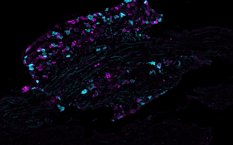
CREDITS: Max Delbrück Center/Sampurna Chakrabarti