Without studies on animals and their basic biological functions, scientists would not be able to make the discoveries that lead to the development of the life-saving drugs and treatments that hit the headlines. So, what animals are used in basic research and how near are we to seeing them replaced by non-animal methods?
A significant part of the use of animals in research is in what is called basic or fundamental research, where scientists seek to understand the crucial processes that govern life without an immediate aim to develop a specific treatment.
Cell biology, which is the study of how cells function, grow and interact, aims to understand life at the microscopic level without necessarily having an immediate medical application. But this basic knowledge has been critical in driving major advances in translational science.
For example, discoveries in cell biology revealed how cells divide, how they communicate and how they die—processes that underpin cancer, immune responses and tissue regeneration. These insights have led directly to the development of targeted cancer therapies, vaccines and stem cell treatments. Direct research of these mechanisms in humans is often impractical and unethical, because it typically involves invasive procedures, manipulation of genes and protein levels, unpredictable risks, not having an immediate therapeutic benefit and making it difficult to ensure truly informed, voluntary consent.
Basic research focuses on understanding fundamental biological processes and mechanisms without immediate practical application. In contrast, translational research builds on this knowledge to develop new therapies, diagnostics or medical technologies that can directly improve human and animal health.
Scientist who studies how the body develops before birth (embryonic development) defines basic research as:
the basics of all new knowledge that can eventually be applied to solve problems of humanity. This is the main power of fundamental knowledge, because you can find something by chance that you didn’t even know existed.
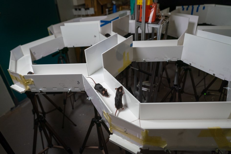
Mice exploring maze for the first time accompanied by cagemates. CREDIT: Radboud University
Animals are essential in basic research because they allow scientists to investigate the core mechanisms of life using techniques that cannot be ethically or practically performed on humans. Researchers employ methods such as genetic editing to manipulate genes and observe how these changes affect cellular processes, development and disease progression. Researchers also use drugs to trigger specific conditions, and advanced imaging and assays to monitor how organs and body systems function. These methods—applied to living organisms ranging from fruit flies and zebrafish to mice and monkeys—provide a controlled environment to study everything from how cells communicate to the origins of complex diseases.
Genetically altered (GA) animals—including mice, zebrafish, rats, monkeys and fruit flies—are indispensable tools in basic research. Modern techniques of gene editing, such as CRISPR/Cas9, allow scientists to precisely manipulate gene function in specific cell types, making it possible to turn genes “on” or “off” and change or add genes in a cell. This approach also provides a controlled experimental setting to recreate human genetic diseases, enabling researchers to explore underlying biological mechanisms. GA animals have been particularly effective in advancing our understanding of proteins and molecules responsible for biological processes such as development and ageing, but also of diseases like cystic fibrosis, cancer and neurodegenerative disorders like Alzheimer’s and Huntington’s disease. GA mice have also been critical for deciphering the function of different types of brain cells, while ‘humanised’ animals—where human genes are introduced into an animal’s genome—enhance the predictive value of preclinical studies. For example, GA mice engineered to express human polio receptors now serve as reliable alternatives to primates in vaccine safety testing, reflecting the growing role of these mice in translating basic research into medical advancements.
Despite their value, the creation and breeding of GA animals is a complex, costly and ethically sensitive process. Current genetic modification technologies don’t always work the same way in every animal, so the intended change may not appear in all GA animals. This leads to a surplus of animals that are bred but not used in experiments. To address this, strict regulatory frameworks and the 3Rs (Replace, Reduce, Refine) are applied, aiming to minimise animal use and suffering while continuing to refine these procedures. As gene editing technologies advance, the precision of genetic alterations is expected to improve further, reducing surplus and enhancing the relevance of GA animals in both basic and translational research.
Beyond gene editing, basic research using animals uses various techniques to explore how biological systems function. Advanced imaging methods, such as mini microscopes, allow scientists to track brain activity in living animals, while techniques that measure electrical signals of cells or stimulate specific cells, like those used to study brain cell communication, help researchers understand brain function and diseases. Additionally, large-scale analyses of genes, proteins and metabolism provide a deeper look at how cells operate and respond to different conditions. Recently, US scientists developed a method that makes the mouse scalp transparent, enabling researchers to observe brain development in real time and track how neural circuits form in response to different stimuli.
The sequencing of the mouse genome in 2002 revealed that mice share 99% of their genes with humans. This genetic similarity has established mice as invaluable models for studying how diseases and genes function. Their widespread use in basic research has deepened our understanding of basic biological processes, contributing to the development of animals that can mimic diseases – by removing or introducing genes that are responsible for a condition, as mentioned above – and in this way leading to drug discovery.
Discoveries from basic research
Embryonic development
How embryos develop before birth – so-called embryonic development – is a critical process that determines the health of the resulting foetus and may also have significant health repercussions into adulthood. Thus, although a mouse with two extra hind limbs where its genitals should be sounds alarming, the study of such specimens in fact reveals the factors that influence embryonic development.
Here, a groundbreaking genetic experiment by EARA member the Gulbenkian Institute for Molecular Medicine (GIMM), Portugal, made the unexpected discovery that such body malformations resulted from a change in the structure of the embryo’s DNA, leading to extra limbs instead of genitalia, and providing a new understanding of how genes guide the formation of such complex body structures. Without animal research, this breakthrough might have gone unnoticed, and, as a result, researchers may not have known how changes in the DNA structure impact cell function – knowledge that has the potential to influence fields like cancer research and immune function.
Also in developmental biology, zebrafish have provided crucial knowledge about eye development and function. Researchers at EARA members the GIMM, Portugal, and the Max Planck Institute, Germany, uncovered a new process in retina formation, where nerve cells adjust their position to allow other cells to divide and build the retina. In Belgium, studies at EARA members KU Leuven and Université Libre de Bruxelles showed that adult zebrafish can regenerate damaged retinal nerve cells. These findings could lead to new treatments for vision loss and other brain and eye disorders.
Studies using fruit fly (Drosophila) embryos also revealed how genes control early development. By introducing random alterations in genes, they identified a set of genes—known as homeobox or HOX genes—that guide how an embryo forms different body parts, such as the head, tail and limbs. These studies in fruit flies were complemented by research in other animals, such as mice, where researchers explored how homeobox genes regulate the transformation of a fertilised egg into a fully formed body and how alterations (i.e. mutations) in these genes cause malformations.
Neuroscience
Basic research in neuroscience has allowed scientists to understand how brain cells communicate, leading to major advances in diagnosing and treating brain disorders like Parkinson’s disease and epilepsy.
Zebrafish have contributed to a better understanding of how the brain controls movement. Recent studies in zebrafish identified specific brain cells that are responsible for governing eye and tail movements and for initiating forward movement and steering. These findings help explain how brain circuits coordinate motion—insights that may inform new treatments for motor disorders.
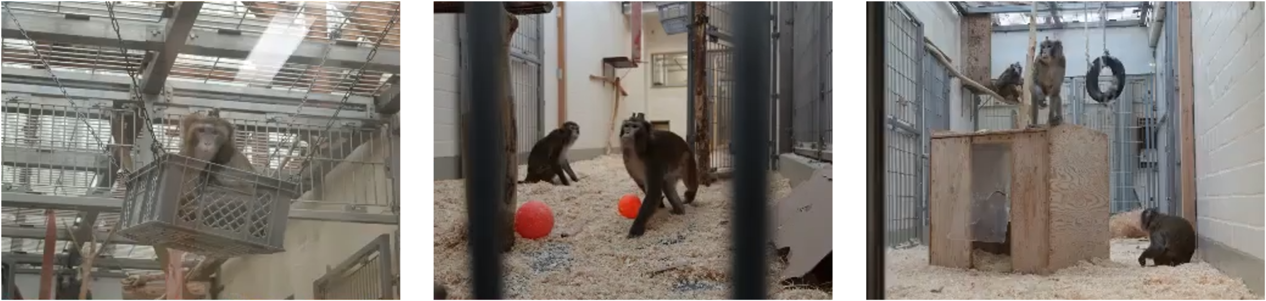
Non-human primates were studied by a team at the University of Bremen, Germany, to uncover how brain cells work during attention tasks. CREDIT: Esperanza Domingo Gil.
Rhesus monkeys play a vital role in basic brain research. At the University of Bremen in Germany, scientists study how the brain supports thinking processes like attention, memory and perception. Their work has helped reveal how the brain's billions of nerve cells form active networks. One part of their research is understanding how different brain cells coordinate to prioritise information during attention tasks, using carefully monitored monkeys that take part in visual tasks – see the EARA Q&A video with Esperanza Domingo Gil, scientist at the University of Bremen in Germany.
My hope is that basic research such as this can unravel these very exciting mysteries of the brain, so that in the future, other researchers can build upon our findings to help heal the brain when disease or age take over.
Esperanza Domingo Gil, scientist and PhD student at the University of Bremen in Germany.
A recent achievement in neuroscience was the creation of the first complete map of the brain of a fruit fly, which is smaller than a poppy seed but has over 140,000 neurons establishing billions of connections. By creating a detailed map of a fully functioning brain, researchers from Princeton University in the US, along with EARA members Freie Universität Berlin and Johannes Gutenberg-Universität Mainz in Germany, laid the foundation for understanding how networks of brain cells work.
The evolution of deep brain stimulation (DBS) as a treatment for Parkinson’s disease (PD) exemplifies basic research studies in animals that paved the way for today’s highly refined therapies. Initial studies in monkeys discovered the network ofbrain cells responsible for movement, and in 1986 researchers identified several distinct networks associated with different brain functions – including those that control movement and eye positioning – and described complex pathways that shape movement. These eventually became the basis for understanding movement disorders.
In the clinical setting, early approaches to treat PD-related tremors involved a procedure called thalamotomy, where electrical stimulation is applied to permanently destroy the brain regions responsible for the tremors. Inspired by observing the temporary benefits of electrical stimulation, researchers then explored a more sustained, less invasive approach, leading to the development of DBS as a long-term therapy for movement disorders. Monkey studies further validated DBS’s effectiveness when applied to animals mimicking human Parkinson’s symptoms, showing that this strategy was able to tackle issues with movement, with fewer side effects than traditional electrical stimulation procedures.
Today, DBS has become a crucial therapy to reduce muscle tremors and manage symptoms in thousands of Parkinson’s patients. Many patients describe DBS in very positive ways, saying it changed their lives or made life easier. For example, a patient who had tremors, painful cramps and uncontrollable movements before DBS described the treatment as, “unbelievably wonderful, it feels like a new life”.
DBS is also being studied as a treatment for weakened muscle strength caused by stroke and traumatic brain injury, showing a promising improvement in the arm and hand function in human patients.
Basic research on the vagus nerve – one of the longest and most important nerves that controls heart rate, breathing and digestion – led to the development of treatments that regulate nerve signals and control brain activity – neuromodulation. These treatments were among the first used to treat epilepsy. Early studies in cats and monkeys revealed that vagus nerve stimulation (VNS) could change brain activity. Building on these initial works, Zabara in 1985 was the first to show that VNS reduced seizures in dogs, providing the crucial proof-of-concept that led to the development of VNS therapy for drug-resistant epilepsy. Over two decades of studies in humans have confirmed that VNS can reduce seizure frequency by at least 50% in roughly half of treated patients after two years.
A study at the University of California in the US looked at a protein called klotho, whose levels are naturally decreased as we age. In 1997, researchers found mice with a genetic change that disrupted the klotho gene. These mice showed symptoms resembling human ageing—such as infertility, osteoporosis and shorter lifespans. When scientists replenished the levels of the klotho protein in these mice, many symptoms improved, suggesting the role of this protein in regulating processes related to ageing. Later, older monkeys treated with klotho also did better in memory tests. This research suggests that klotho might help protect brain function and could be important for treating brain diseases like Alzheimer’s and Parkinson’s.
“Given the close genetic and physiological parallels between primates and humans, this could suggest potential applications for treating human cognitive disorders.” – Marc Busche, Neurologist at University College London., Neurologist at University College London., Neurologist at University College London.
Parkinson's disease patient explains how deep brain stimulation works and the impact that it had on his life. CREDIT: Understanding Animal Research.
Immunology
The immune system is complex and multifaceted, existing in nearly all organisms, acting as the major line of defence against infections and illnesses through a diverse range of processes and molecules. Basic research in immunology has revealed how the immune system detects and fights infections, paving the way for vaccines, allergy treatments and cancer immunotherapies.
In 2024, the Albert Lasker Basic Medical Research Award recognised Zhijian “James” Chen at UT Southwestern Medical Center, USA, for a landmark discovery in immunology: a specific protein (cGAS) that detects bacteria inside the cells and triggers immune defences. Further research showed that this protein also plays a role in autoimmune diseases and studies in mice revealed that changes in this protein can result in fatal autoimmune conditions. Thus, the initial discovery of the function of cGAS provided a pathway toward new treatments for autoimmune diseases.
By studying mice, researchers at EARA member University of Liège in Belgium discovered a new type of immune cells that can regenerate the lungs after viral infections. These immune cells were also found in patients with suspected pneumonia and mice genetically modified to lack them had reduced lung recovery after infection.
Mice also played a crucial role in understanding the microbiome – the collection of all microbes, such as bacteria, fungi and viruses, that naturally populate the human body. Building on this knowledge, a recent study led by the University of Cambridge, UK, showed that altering the gut microbiome of pregnant mice led to significant changes in the brain development of their offspring – suggesting that by providing beneficial bacteria to the mother, it might be possible to enhance foetal growth and neurological development.
This exciting discovery may pave the way for future clinical studies that explore the critical role of the maternal microbiome in supporting healthy brain development before birth.
Another key biological process studied using mice is how the body controls involuntary reflexes like sneezing and coughing upon respiratory infections. US scientists, at Washington University and Georgia Institute of Technology, looked at which brain cells trigger these reactions. They also found that mice genetically modified to lack a specific protein in the nose no longer sneezed when infected with the flu virus. In a similar experiment, the modification of another protein in the trachea stopped infected mice from coughing, but they could still sneeze. This showed that sneezing and coughing are controlled by separate brain cells.
Decades of studies in animals, such as mice and pigs, allowed scientists to deliver messenger RNA (mRNA) into cells so it stays stable and produces proteins. Early experiments showed that putting mRNA into tiny lipid particles let it safely enter the cells and make proteins that the immune system can detect. This basic research in animals helped create the technology used in today's mRNA vaccines, such as for Covid-19.
Research using mice, ferrets, monkeys, hamsters and pigs—not only for mRNA research but also for studying other coronaviruses such as MERS— also helped speed up the development of Covid-19 mRNA vaccines. Testing the vaccines in different animal species was instrumental in ensuring they were safe and effective before proceeding to studies in humans. Moreover, the continuity of vaccine research—such as the Oxford/AstraZeneca vaccine for other coronaviruses—allowed scientists to quickly use its results to improve Covid-19 vaccines.
The studies on how mRNA works didn’t just contribute to the rapid development of Covid-19 vaccines but also paved the way for new treatments and vaccines for other diseases. For instance, in a study at University College London and King’s College London in the UK, a therapy based on the same mRNA technology used in Covid-19 vaccines successfully corrected a rare liver disease, argininosuccinic aciduria, in mice, demonstrating the potential to treat other rare diseases. Scientists have also developed an effective vaccine against malaria, using the mRNA technology first seen in the successful Covid-19 vaccines.
Messenger RNA has revolutionised the field of vaccines during the Covid-19 pandemic. We believe it can now do the same for rare diseases.
Julien Baruteau, at UCL Great Ormond Street Institute of Child Health
Researchers at EARA member Biomedical Primate Research Centre in the Netherlands and University of Tennessee Health Science Center in the US, studied macaques infected with simian immunodeficiency virus (SIV), a close relative of HIV, to study how the virus can affect the brain. In this case, the study uncovered underlying biological mechanisms without the immediate goal of developing treatments. Scientists observed not only that macaques with increased viral loads of SIV exhibited neurological symptoms, but they also had proteins from bacteria, which are normally absent in a healthy brain, likely contributing to increased inflammation and neurological damage.
Zebrafish are also important animals for studying the immune system. US researchers at the University of Wisconsin–Madison used transparent zebrafish to watch immune cells travel through the body in real time. This allowed them to confirm, for the first time, long-standing theories about how these cells travel in the body.
Fruit flies also have a complex immune system, allowing researchers to study how macrophages – immune cells responsible for absorbing harmful pathogens – penetrate tissues, thus informing future research into immune response, wound healing and inflammatory diseases.
Another example of basic research is the 1956 study by Bruce Glick on chickens’ immune systems, which was initially considered unclear but was awarded the Golden Goose Award in 2018. Glick’s research discovered that removing the bursa of Fabricius – an organ unique to birds – prevented chickens from producing antibodies – proteins from the immune system that help the body fight infections. Although its practical value wasn’t immediately clear, this finding has become foundational in immunology, paving the way for vaccines, treatments for autoimmune diseases and cancer.
Cancer research
The identification of proteins with dual roles is not something new in molecular biology research. For instance, the important role of the SOX17 gene in the development of the pancreas, liver and biliary (bile) system was first identified in mice when studying what impacts tissue differentiation and organ development. This research revealed that SOX17 contributes to the formation of key organs by regulating how cells transform into specific tissue types and how changes in SOX17normal levels result in severe abnormalities in how the embryo develops. However, the story of SOX17 doesn't stop at development. In recent years, researchers have turned their attention to its role in cancer: SOX17 also interferes with how cancer cells avoid detection by the immune system in colorectal cancer. By diminishing the immune response, SOX17 allows early-stage cancer cells to go undetected and continue their dangerous transformation. This discovery suggests that SOX17 could not only serve to detect early colorectal cancer but might also emerge as a novel therapeutic target.
Biology and ecology
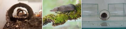
Animals also play an important role in biology and ecology research. Investigations into topics like animal behaviour, species interactions, population dynamics and evolutionary processes, such as hibernation and the effects of environmental changes, require observing and experimenting on living organisms in natural or controlled settings. These complex, whole-animal processes are not possible to capture in lab-grown cells alone. Wild species, such as small mammals, are also used to provide insights into ecology (the relationship between organisms and their environment), conservation and environmental monitoring. For example, EARA member the University of Lisbon (ULisboa) in Portugal studies wild animals, including shrews, voles and wood mice. These animals are brought into the animal facilities for short periods of time to understand how urban development affects wildlife. Another ULisboa project looks at how being in captivity may change animals’ behaviour so that research findings still apply to animals in the wild.
Examples of wild-caught small mammals (left: Microtus lusitanicus; centre: Crocidura russula; right: Microtus duodecimcostatus) used in ecology research to understand the impact of environmental changes on wildlife. CREDIT: Ana Cerveira and MaÃlis Carrilho, University of Lisbon, Portugal.
Other fields
Mice have also contributed to the understanding of the gastrointestinal system. In Germany, researchers at University of Würzburg with EARA members Charité Berlin and Max Planck Institute for Infection Biology, studied the development of the gastroesophageal junction—the connection between the oesophagus and the stomach. By following how individual cells grow from the embryo to adulthood in mice, and using lab-grown organoids derived from mouse and human tissues, researchers discovered how this junction forms.
One important discovery from research on zebrafish was finding new components of the heart’s nervous system. Scientists used to think that the heart rate was controlled only by automatic signals from the brain. But a study in zebrafish showed that the heart also has its own network of nerve cells that help control the heartbeat. Researchers at EARA member the Karolinska Institute in Sweden and at Columbia University in the US mapped these nerve cells in zebrafish hearts, revealing how complex they are. This has changed how scientists understand heart function and may lead to new treatments for heart disease.
We were surprised to see how complex the nervous system within the heart is.
Konstantinos Ampatzis, senior researcher at the Karolinska Institute
Studies in fruit flies allowed scientists to understand how insulin-producing cells in the brain respond to movement. Researchers at the University of Würzburg found that when flies were active, insulin activity dropped to release energy, and when they stopped, insulin levels rose again to restore energy reserves. These findings into insulin regulation help explain how the body manages energy and could improve our understanding of metabolic disorders like diabetes and how exercise affects blood sugar levels.
Which animals are used in basic research?
Basic research uses different animal species depending on the scientific question being asked. No single animal model can answer all questions about biology or disease, so researchers select species whose biology best matches the process under study. Whenever possible, simpler or smaller organisms are used, and more complex animals are only involved when no other model can provide the necessary information.
Recent research has mapped the complete set of zebrafish genes (whole genome), revealing how many genes zebrafish and humans share. In 2013, the zebrafish genome project conducted by the Wellcome Sanger Institute, UK, found that 70% of human genes have at least one zebrafish counterpart, and that 84% of genes linked to human disease are represented in zebrafish. Zebrafish are particularly valuable in basic research because their embryos are transparent, allowing scientists to directly observe how organs and tissues form and how disease affects the body during early development. They also develop rapidly and produce large numbers of embryos, making them useful for studying biological processes efficiently and, in many cases, reducing the need for using mammals.
Rodents such as mice and rats are the most widely used mammals in basic research because they share many biological and genetic similarities with humans. Around 95% of mouse genes have a human counterpart, allowing researchers to study how specific genes can influence biological functions like development, digestion, metabolism, memory and immunity. The mouse genome, meaning its complete set of DNA, has been fully mapped, which allows researchers to switch genes on or off to understand their role in health and disease. Genetically altered (GA) mice are therefore particularly valuable for mimicking human and veterinarian diseases. Mice have a relatively short lifespan of two to three years and reproduce quickly, enabling researchers to observe disease progression and treatment effects over a full life course in a shorter period than in humans. Their rapid reproduction and large litters support studies across generations, which is especially useful for research on ageing, vaccines and hereditary conditions. Their small size means they require less space and fewer resources compared with larger mammals, and many procedures can be refined to minimise discomfort. In some studies, mice can also replace larger animals entirely. While mice are commonly used for genetic and molecular studies, rats are often preferred for research on behaviour, cardiovascular function and complex surgical procedures. Their larger size and well-characterised physiology make them particularly suitable for studies requiring detailed monitoring or physical interventions.
A small number of NHPs (e.g. monkeys) are used in basic research and translational research, primarily in studies of the nervous system and infectious diseases. Their use is strictly regulated and only approved when no other animal or non-animal method can answer the research question. Non-human primates are used in limited cases because they share specific aspects of brain organisation, immune responses and metabolism with humans that cannot be fully replicated in other species. Some conditions, such as atherosclerosis, osteoporosis and hypertension, occur naturally in primates, making them particularly relevant for understanding these diseases.
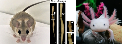
On the left, the African spiny mouse, Acomys, and his unique regenerative ability to fully heal a spinal cord transection. Credits: Joana Nogueira-Rodrigues et al./Developmental Cell. On the right is the axolotl (Ambystoma mexicanum), an aquatic salamander, that can regenerate limbs and complex tissues. Credits: Kramer et al./Disease Models and Mechanisms.
Studying how unusual animals like the African spiny mouse (Acomys) and axolotl change over time and adapt to their environments is driving breakthroughs in regenerative medicine, including therapies for spinal cord injury (SCI). Unlike most mammals, the spiny mouse can regenerate its skin, hair follicles and cartilage after injuries, allowing it to heal without scarring. Scientists found that these animals change how proteins are present at the injury site, creating a scar-free environment that encourages new cell growth and recovery. Researchers from EARA member i3s in Portugal are developing SCI treatments that are based on this regenerative capacity of the African spiny mouse, which can fully regenerate nervous tissue even after severe SCI – an ability unmatched in any other adult vertebrate.
"This work goes far beyond demonstrating this regenerative capacity in Acomys, as we show the molecular mechanisms underlying this phenomenon (...) Until now, advances had been made in understanding the function of certain proteins that aid in the regenerative process. But Acomys has allowed us to see that we need to look at the signature of the sugars present and their biosynthesis, which will elevate the work being done in this field." – Mónica Sousa, scientist at i3S in Portugal.
Similarly, the axolotl (Ambystoma mexicanum), an aquatic salamander, can regenerate entire limbs and complex tissues. These animals have evolved and developed characteristics that help them escape predators and survive in nature and have inspired scientists to explore ways of replicating similar processes in humans.
Limitations of animal models for basic research
Animal research remains irreplaceable in basic research, especially when studying complex biological phenomena that we cannot yet mimic accurately in the lab or with computational methods. However, the use of animals in basic research does come with some inherent limitations, since biological, genetic and physiological differences between humans and animals mean that findings in one species do not always translate directly to another. These differences require caution from both an ethical and scientific standpoint, as well as a commitment to minimising animal use by exploring alternative methods.
Yet, in cases that involve the study of intricate biological processes that result from the interaction between complex organ systems, where no other model can replicate the biological process so closely, animal research remains the only viable option. Moreover, we cannot model what we don’t yet understand—a challenge that is especially true for complex organs like the brain, where basic studies in animals remain essential to uncovering the neural mechanisms that underlie human cognition, emotion and disease. The balance lies in understanding when animal research is essential for exploring unknown biological mechanisms, all while advancing methods to ensure animal welfare and seeking alternatives when possible.
It’s important for scientists to not make exaggerated promises in their research and they should emphasise the importance of the long-term acquisition of knowledge in basic research using animals.
Gilles Laurent, scientist at the Max Planck Institute for Brain Research, Germany.
New approach methodologies in basic research
New approach methodologies (NAMs) are essential tools in basic research, as well as in regulatory safety testing, providing insights into biological processes that are important to understanding life’s complexity. Replacement techniques, such as cell cultures and computational models, offer valuable alternatives that allow scientists to explore cellular processes, genes and how cells grow and form tissues, without relying solely on animals.
Similarly, methods like advanced imaging, innovative biomaterials, organ-on-a-chip technologies and organoids allow researchers to analyse cellular and tissue interactions in a controlled, in vitro environment in the lab. Organoids, which mimic some of the function of entire organs, have opened new avenues for studying how cells differentiate, as well as to model diseases and organ development. In neuroscience, brain organoids provide a three-dimensional model for exploring brain cell networks and different brain signals, which are challenging to replicate in single cell cultures in the lab. Computer models and simulations, meanwhile, enable testing of predictions about the processes happening in cells, such as metabolism, the blood-brain barrier, and cell communication – and even allow information to be collected from animal studies in a completely non-invasive way. Certain aspects of the human brain, immune system or infectious diseases cannot be studied in a petri dish or computer model with the necessary level of detail.
Considerable advances have been made in developing (non-animal) alternatives, but animal models remain unavoidable at the moment to understand some more complex biological or physiological processes involved in health, disease and biodiversity.
In vitro and computer models allow researchers to conduct detailed investigations into protein functions, gene regulation and molecular interactions with a high level of precision and control. For example, scientists are using cell-based high-throughput screening with thousands of small molecules, which manipulate specific genes or proteins, to identify previously unknown regulators of processes involved in diseases and development. These insights would be difficult, impractical or ethically challenging to obtain in animals due to the high number of samples required for high-throughput screening. Additionally, the precise control over experimental conditions achievable in cell-based systems is much harder to replicate in complex animal organisms, where confounding effects, such as fluctuations in hormone levels, can make it difficult to interpret the data correctly.
While in vitro and computational methods can't fully replicate the complexity of an entire organism, they are invaluable in building a deep understanding of life's basic mechanisms. These tools enable researchers to refine hypotheses, make precise adjustments and validate early findings, laying a solid foundation before advancing to animal models. Scientists can then focus animal research on studying the dynamic and complex responses of biological organisms.
The key is to find the right balance – leveraging animal research where it is truly essential for making groundbreaking discoveries, especially when studying the unknown, while continuously developing strategies for replacement, reduction and refinement. By doing so, researchers can commit to ethical priorities while also unlocking the critical insights that underpin the next generation of life-changing innovations.For the foreseeable future, the ethical and careful use of non-human primates will remain essential to advancing safe solutions to human and animal diseases.